Home
/ Diagram Of Liver Cell / Liver Cell With Labelled Structures Illustration Stock Image C043 6871 Science Photo Library - Example of blood, neurons, cardiac, bone, intestinal, epithelial, fat, liver and.
Diagram Of Liver Cell / Liver Cell With Labelled Structures Illustration Stock Image C043 6871 Science Photo Library - Example of blood, neurons, cardiac, bone, intestinal, epithelial, fat, liver and.
Diagram Of Liver Cell / Liver Cell With Labelled Structures Illustration Stock Image C043 6871 Science Photo Library - Example of blood, neurons, cardiac, bone, intestinal, epithelial, fat, liver and.. Smartdraw includes 1000s of professional healthcare and anatomy chart templates that you can modify and make your own. Human anatomy detailed diagram of various human organs liver, heart, kidneys, lungs, colon, intestine, stomach, brains, etc can be used in. Whatever an organism does for survival it does for the survival of its cells. Form specific compounds such as coagulation factors and. The liver is responsible for the production of several vital protein components of blood plasma:
The incidence of liver diseases is rising and there are limited treatment options. Co₂ from cellular respiration is excreted from the lungs. Two diagrams of liver structure removed for copyright reasons. Hepatocyte nuclei often contain a prominent nucleolus. The cell is the fundamental unit of life.
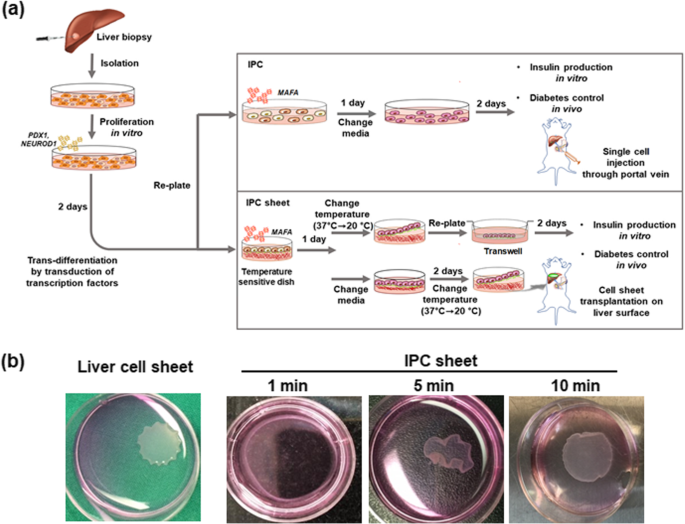
Improvement Of The Therapeutic Capacity Of Insulin Producing Cells Trans Differentiated From Human Liver Cells Using Engineered Cell Sheet Springerlink from media.springernature.com Its other roles in metabolism. The human liver is an essential multifunctional organ. The cell is the fundamental unit of life. Liver sinusoidal endothelial cells (lsecs) act as a filter between the lumen of the hepatic sinusoids and the surrounding hepatocytes. Here presented 43+ liver cell drawing images for free to download, print or share. Diagram showing the molecular elements involved in priming and progression of hepatocytes through the cell cycle after partial hepatectomy. They are liver cells with a large nucleus, a prominent golgi and ↑ mitochondria. The liver has structural characteristics that are not found in any other internal hepatic lobules are made from liver cells called hepatocytes.
Hepatic stellate cells (hscs) are specialized liver pericytes residing in the space of disse lining hepatocytes and endothelial sinusoidal cells.
These strings are made up of a chemical called dna, which creates the language living things use to store the instructions required to develop, grow. Ƽ intricately involved in carbohydrate, fat, and protein metabolism. It is a large organ, with its major lobe occupying the right side of the abdomen below the diaphragm, while the narrower left lobe extends all the way across the abdomen to the left. How are bile pigments formed and excreted? Human anatomy detailed diagram of various human organs liver, heart, kidneys, lungs, colon, intestine, stomach, brains, etc can be used in. Hepatic stellate cells (hscs) are specialized liver pericytes residing in the space of disse lining hepatocytes and endothelial sinusoidal cells. Two diagrams of liver structure removed for copyright reasons. Prothrombin and fibrinogen proteins are coagulation factors involved in the formation of blood clots. Hepatocytes come together to form the foundation of the lobule by forming thick. What your do and liver functions that are essential to life. The stellate fat storing cell. Smartdraw includes 1000s of professional healthcare and anatomy chart templates that you can modify and make your own. Blood flows through the liver sinusoids and empties into the central vein of each the kupffer cells of liver are phagocytic cells, helps in phagocytosis of dead blood cells and bacteria from the blood.48.
7710x4991 liver cell diagram liver histology labpedia. 2.3.2 annotate the diagram from 2.3.1 with the functions of each named structure. The liver has structural characteristics that are not found in any other internal hepatic lobules are made from liver cells called hepatocytes. Documents similar to liver pathophysiology and schematic diagram. Ƽ store vitamins and minerals;
Histolab Part 19 from www.meddean.luc.edu Learn about the human liver. The liver is a vital organ found in humans and other vertebrates. Albumins are proteins that maintain the isotonic environment. Human anatomy detailed diagram of various human organs liver, heart, kidneys, lungs, colon, intestine, stomach, brains, etc can be used in. Below is a diagram of a compound light microscope. A diagram of the liver, pancreas, and bile passage. It is a large organ, with its major lobe occupying the right side of the abdomen below the diaphragm, while the narrower left lobe extends all the way across the abdomen to the left. Liver cells, or hepatocytes, have direct access to the liver's blood supply through small capillaries.
Below is a diagram of a compound light microscope.
However, the cellular composition of the liver remains poorly understood. Another type of liver cell is the endothelial cells. Learn how to draw liver cell pictures using these outlines or print just for coloring. 7710x4991 liver cell diagram liver histology labpedia. Co₂ from cellular respiration is excreted from the lungs. Blood flows through the liver sinusoids and empties into the central vein of each the kupffer cells of liver are phagocytic cells, helps in phagocytosis of dead blood cells and bacteria from the blood.48. In a healthy liver, hscs maintain extracellular matrix (ecm) homeostasis and accumulate vitamin a in the form of retinyl esters in cytoplasmic lipid droplets. There are 4 basic cell types that reside in the liver: A diagram of the liver, pancreas, and bile passage. Ƽ intricately involved in carbohydrate, fat, and protein metabolism. What your do and liver functions that are essential to life. Documents similar to liver pathophysiology and schematic diagram. Hepatocytes are polygonal epithelial cells with abundant eosinophilic, granular cytoplasm and large, centrally located round nuclei.
Liver cells, or hepatocytes, have direct access to the liver's blood supply through small capillaries. There are 4 basic cell types that reside in the liver: The liver has structural characteristics that are not found in any other internal hepatic lobules are made from liver cells called hepatocytes. Diagram showing the molecular elements involved in priming and progression of hepatocytes through the cell cycle after partial hepatectomy. 2.3.1 draw and label a diagram of the ultrastructure of a liver cell as an example of an animal cell.
Katp Channels Regulate Mitogenically Induced Proliferation In Primary Rat Hepatocytes And Human Liver Cell Lines Journal Of Biological Chemistry from els-jbs-prod-cdn.jbs.elsevierhealth.com Learn how to draw liver cell pictures using these outlines or print just for coloring. Blood flows through the liver sinusoids and empties into the central vein of each the kupffer cells of liver are phagocytic cells, helps in phagocytosis of dead blood cells and bacteria from the blood.48. It is a large organ, with its major lobe occupying the right side of the abdomen below the diaphragm, while the narrower left lobe extends all the way across the abdomen to the left. There are 4 basic cell types that reside in the liver: 7710x4991 liver cell diagram liver histology labpedia. The liver parenchyma is primarily comprised of hepatocytes. Albumins are proteins that maintain the isotonic environment. Medical labeled diagram with all kind cells.
Prothrombin and fibrinogen proteins are coagulation factors involved in the formation of blood clots.
2.3.1 draw and label a diagram of the ultrastructure of a liver cell as an example of an animal cell. The liver cells have two different sources of blood supply. Ƽ intricately involved in carbohydrate, fat, and protein metabolism. What your do and liver functions that are essential to life. The stellate fat storing cell. Example of blood, neurons, cardiac, bone, intestinal, epithelial, fat, liver and. 2.3.2 annotate the diagram from 2.3.1 with the functions of each named structure. The liver is partially surrounded by the ribs, and extends from the level of the fifth intercostal space to the lower margin of the right rib cage, which protects this highly vascular organ. Liver sinusoidal endothelial cells (lsecs) act as a filter between the lumen of the hepatic sinusoids and the surrounding hepatocytes. Whatever an organism does for survival it does for the survival of its cells. 2.3.1 draw and label a diagram of the ultrastructure of a liver cell as an example of an animal cell. The human liver is an essential multifunctional organ. Schematic diagram showing influence of ha on angiogenesis in liver ecs.
Below is a diagram of a compound light microscope diagram of liver. Hepatic stellate cells (hscs) are specialized liver pericytes residing in the space of disse lining hepatocytes and endothelial sinusoidal cells.

