Home
/ Brain Anatomy Map : Print Map Quiz Internal External Brain Functions Hotspot Brain Brain Anatomy Life Science - Together, the brain and spinal cord that extends from it make up the central nervous system, or cns.
Brain Anatomy Map : Print Map Quiz Internal External Brain Functions Hotspot Brain Brain Anatomy Life Science - Together, the brain and spinal cord that extends from it make up the central nervous system, or cns.
Brain Anatomy Map : Print Map Quiz Internal External Brain Functions Hotspot Brain Brain Anatomy Life Science - Together, the brain and spinal cord that extends from it make up the central nervous system, or cns.. The human brain is particularly complex and extensive. .anatomy of the brain anatomical chart human brain map rat brain anatomy brain anatomy cerebral cortex labeled brain map cognitive brain map brain sensory map frontal lobe brain. This interactive brain model is powered by the wellcome trust and developed by matt wimsatt and jack simpson; A brain is an organ that serves as the center of the nervous system in all vertebrate and most invertebrate animals. Brain, bones of cranium, sinuses of the face.
Reviewed by john morrison, patrick hof, and edward lein. The human brain is the main central nervous system organ, situated in the head, protected by the cranium. On the left a coronal view of the segments of the middle cerebral artery. New way to browse data. Humans are born with relatively immature brains that continue to develop in size, shape, and structure throughout childhood and adolescence.
Human Brain Anatomical Chart Anatomy Poster Anatomical Poster Anatomy Chart Brain Anatomy Human Brain Brain Poster from i.pinimg.com Structure descriptions were written by levi gadye and alexis wnuk and jane roskams. It serves as a relay station, passing messages back and forth between various parts of the body and the cerebral cortex. The brainstem is the lower extension of the brain, located in front of the cerebellum and connected to the spinal cord. The following table shows the brain regions from the cortex to the brain stem with their functions and associated disorders in english, german and latin. A brain model with arteries and coloured sections, for basic anatomy education. Reviewed by john morrison, patrick hof, and edward lein. What is the brain made of. This interactive brain model is powered by the wellcome trust and developed by matt wimsatt and jack simpson;
The midbrain helps control eye movement and processes visual and.
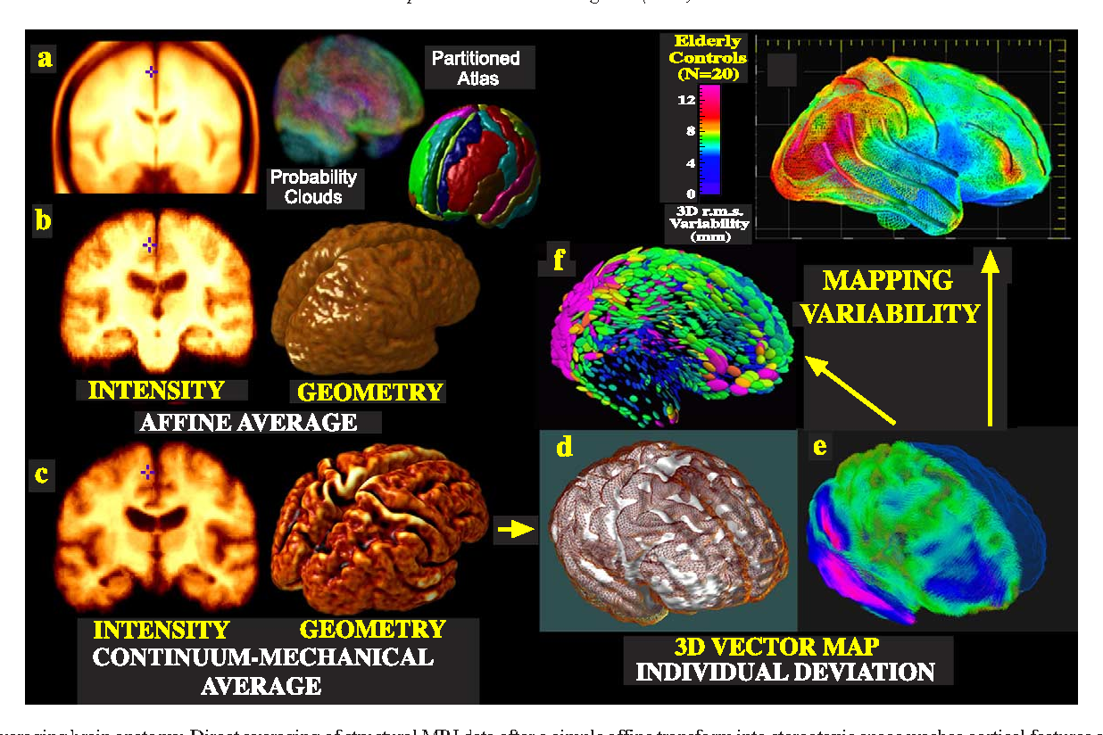
This amazing organ acts as a control center by receiving, interpreting, and directing sensory information throughout the body. Together, the brain and spinal cord that extends from it make up the central nervous system, or cns. The top image shows the four main sections of the cerebral cortex: Humans are born with relatively immature brains that continue to develop in size, shape, and structure throughout childhood and adolescence. Anatomy of the brain there are different ways of dividing the brain anatomically into regions. The mapping effort subdivided each half of the brain into 180 separate partitions, with 83 of those already well established in the field, and 97 new areas ripe for exploration. All references (mainly links to abstracts) are only given in place of many other studies that point towards the same function or disorder. Can you name these brain structures? Foramina, nasal cavity, paranasal sinuses. What is the brain made of. Mapping 'imbalance' in brain anatomy across the lifespan. It consists of three structures: A brain model with arteries and coloured sections, for basic anatomy education.
The brain and spinal cord are the two main structures of the central nervous system. Map of the human brain: Brain magnetic resonance imaging (mri) is a common medical imaging method that allows clinicians to examine the brain's anatomy (1).it uses a magnetic field and radio waves to produce detailed images of the brain and the brainstem to detect various conditions (2).these include tumors, inflammatory ailments, and developmental and structural abnormalities. Map of the human brain: Humans are born with relatively immature brains that continue to develop in size, shape, and structure throughout childhood and adolescence.
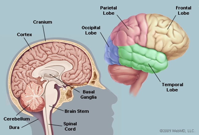
Figure 1 From Mapping Cortical Change In Alzheimer S Disease Brain Development And Schizophrenia Semantic Scholar from d3i71xaburhd42.cloudfront.net A brain is an organ that serves as the center of the nervous system in all vertebrate and most invertebrate animals. There are three major divisions of the brain. It serves as a relay station, passing messages back and forth between various parts of the body and the cerebral cortex. The anatomy of the brain is complex due its intricate structure and function. Let's use a common method and divide the brain into three main regions based on embryonic development: Webmd's brain anatomy page provides a detailed diagram and definition of the brain including its function, parts, and conditions that affect it. Mapping 'imbalance' in brain anatomy across the lifespan. The midbrain, pons and medulla oblongata.
Anatomy of the head on a cranial ct scan :
Anatomy of the head on a cranial ct scan : The allen mouse brain common coordinate framework (ccfv3), a 3d reference atlas, is based on an average of the inherent fluorescence in the. It serves as a relay station, passing messages back and forth between various parts of the body and the cerebral cortex. One new feature is a set of 10 hierarchical nomenclature tables that define and describe all parts of the rat nervous system within the framework of a strictly topographic system devised previously for the. The top image shows the four main sections of the cerebral cortex: The anatomy of the brain is complex due its intricate structure and function. Can you name these brain structures? Map of the human brain: In three papers, the potential of using light to unveil the function and anatomy of brain circuits is presented. Thinking, perceiving, planning, and understanding language all lie within the cerebrum's control. Utilize the model of the human brain to locate the following structures / landmarks for the Anatomy of the brain there are different ways of dividing the brain anatomically into regions. On the left a coronal view of the segments of the middle cerebral artery.
Webmd's brain anatomy page provides a detailed diagram and definition of the brain including its function, parts, and conditions that affect it. Dural venous sinuses, veins, arteries. While there is still a great deal that researchers do not yet know about the brain, they have learned a great deal about the anatomy and function of the brain. A brain model with arteries and coloured sections, for basic anatomy education. Foramina, nasal cavity, paranasal sinuses.
Brain Human Anatomy Picture Function Parts Conditions And More from img.webmd.com All references (mainly links to abstracts) are only given in place of many other studies that point towards the same function or disorder. The brain is a complex organ that controls thought, memory, emotion, touch, motor skills, vision, breathing, temperature, hunger and every process that regulates our body. Structure descriptions were written by levi gadye and alexis wnuk and jane roskams. Brain, bones of cranium, sinuses of the face. Dural venous sinuses, veins, arteries. The lowest part of the brainstem, the medulla is the most vital part of the entire brain and contains important control centers for the heart and. Map of the human brain: Reviewed by john morrison, patrick hof, and edward lein.
The cerebrum, the largest part of the human brain, is associated with higher order functioning, including the control of voluntary behavior.
Let's use a common method and divide the brain into three main regions based on embryonic development: Anatomy of the head on a cranial ct scan : In three papers, the potential of using light to unveil the function and anatomy of brain circuits is presented. Thinking, perceiving, planning, and understanding language all lie within the cerebrum's control. The midbrain helps control eye movement and processes visual and. The midbrain, pons and medulla oblongata. It consists of three major parts: Brain map anatomy helps medical students change the way they make notes, prioritize information, improve their memory in learning anatomy. It consists of three structures: Webmd's brain anatomy page provides a detailed diagram and definition of the brain including its function, parts, and conditions that affect it. Mapping 'imbalance' in brain anatomy across the lifespan. It embodies 2% of body mass, but it takes approximately. All references (mainly links to abstracts) are only given in place of many other studies that point towards the same function or disorder.
Thinking, perceiving, planning, and understanding language all lie within the cerebrum's control anatomy map. It serves as a relay station, passing messages back and forth between various parts of the body and the cerebral cortex.
